The way out of the cell changes the protein.
Before a membrane protein can be imaged, it frequently has to be lifted out of the cell membrane. The standard approach uses detergents, which strip away the native lipids associated with the protein. What's left is often an artifact: conformations shift, binding pockets distort, and lipids that were part of the structure simply disappear.
That matters downstream. A drug program built on a distorted structure can spend years and resources designing against a binding site that isn't really there in the living cell.
Prediction doesn't close the gap either. AlphaFold and other AI tools capture sequence-based folding, but they cannot model the native lipid environment, membrane dynamics, or the lipid-dependent conformations a drug actually encounters. For membrane proteins, an experimental structure in a native membrane is still required.
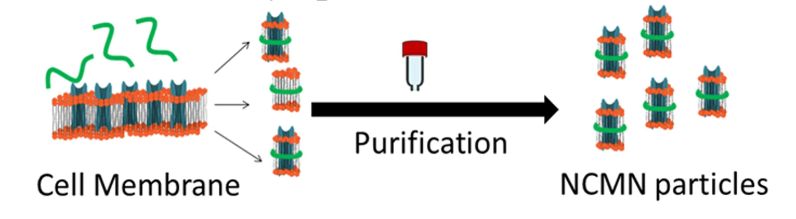
The protein, with its lipids still attached.
The NCMN System extracts a membrane protein together with its associated native lipids as a single stable nanoparticle. No detergents. No engineered mutations, truncations, or fusions to force the protein to behave.
What goes onto the cryo-EM grid is the protein in its real membrane environment, ready to be resolved at high resolution. The result is a structure that reflects the protein as it actually exists in the cell.
Built for structures you can trust.
- Native lipids retained
- No protein engineering required
- Sub-3Å cryo-EM resolution
- Full functional activity preserved
- Homogeneous, reproducible nanoparticles
- Broad target coverage